Optical coherence tomography
OCT creates detailed cross-sectional images of eye tissues, commonly used to examine retinal layers and, when indicated, the optic nerve. The doctor interprets the scan together with symptoms and examination findings.
 Book an appointment
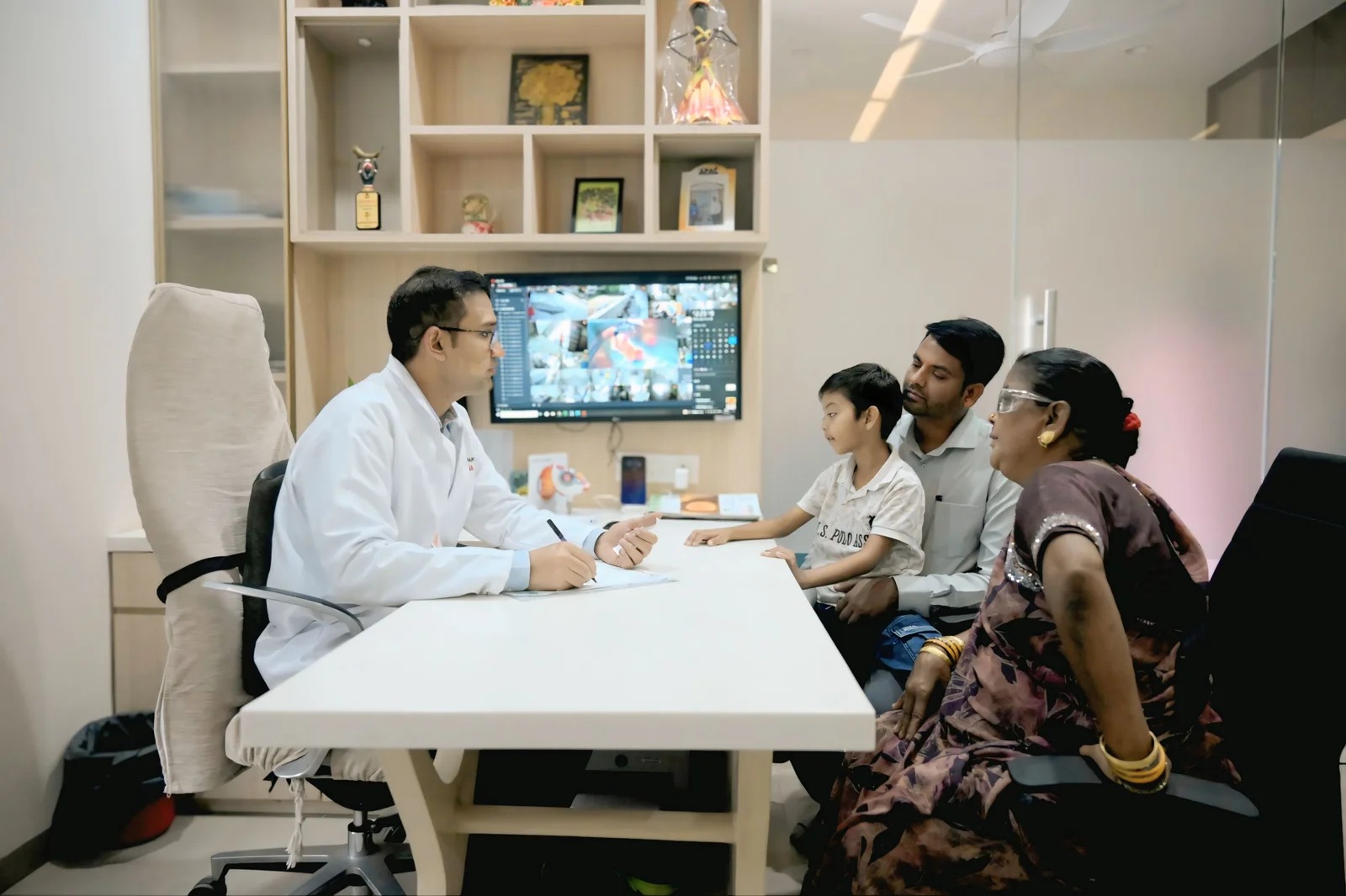
Eye tests · Raipur
Eye investigations can help a clinician assess the retina, eye structure, pressure or focusing. Shree Retina Care’s supplied rate-list material includes OCT, A-scan, B-scan, FFA, pachymetry and fundus photography, among other items. The doctor decides what is appropriate.
The clinic material reviewed does not identify device brands or models. This page does not claim particular equipment specifications or live availability; confirm test names, preparation and current charges with reception.
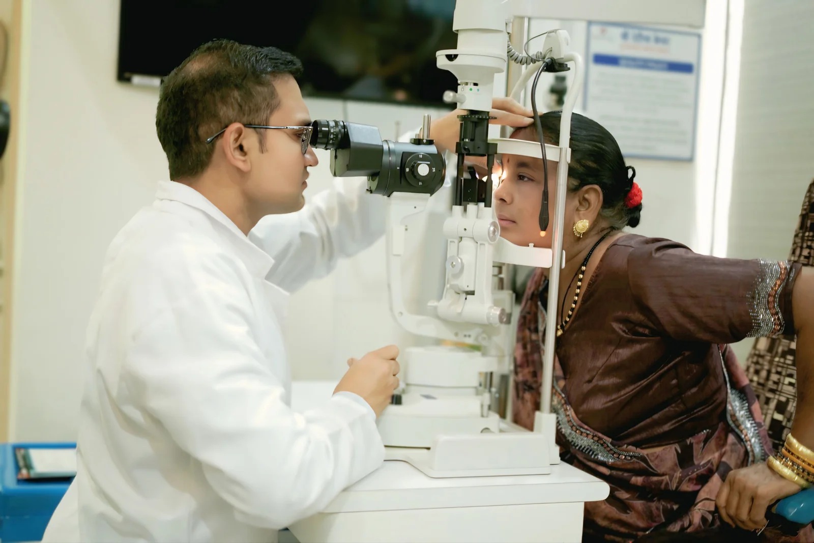
Investigations listed by the clinic
The explanations below are general. A test name does not indicate that every patient needs it, that it is available on a particular day, or that a diagnosis can be made from one test alone.
OCT creates detailed cross-sectional images of eye tissues, commonly used to examine retinal layers and, when indicated, the optic nerve. The doctor interprets the scan together with symptoms and examination findings.
An A-scan uses ultrasound measurements of the eye. It may be used in planning certain eye procedures, including intraocular lens calculations, when the clinician considers it appropriate.
B-scan ultrasound produces an image of structures inside the eye. A clinician may consider it when a direct view of the back of the eye is limited or another question needs assessment.
FFA uses a dye and retinal imaging to assess blood flow in the back of the eye. Ask why it is recommended, what preparation is needed and which risks or after-effects apply to you.
Pachymetry measures corneal thickness. The doctor may use the result as one part of an assessment; it should not be interpreted on its own.
A fundus photograph records an image of the back of the eye. It can help document appearance over time, but the doctor still interprets it in the context of an examination.
Other entries on the supplied list
The clinic-supplied rate-list image also includes FFA, scansys, cyclorefraction, dilation and dry-eye-related testing or charges. The word “Scansys” is not sufficiently specific to explain a machine or test on this page; ask the clinic for the exact test name and purpose.
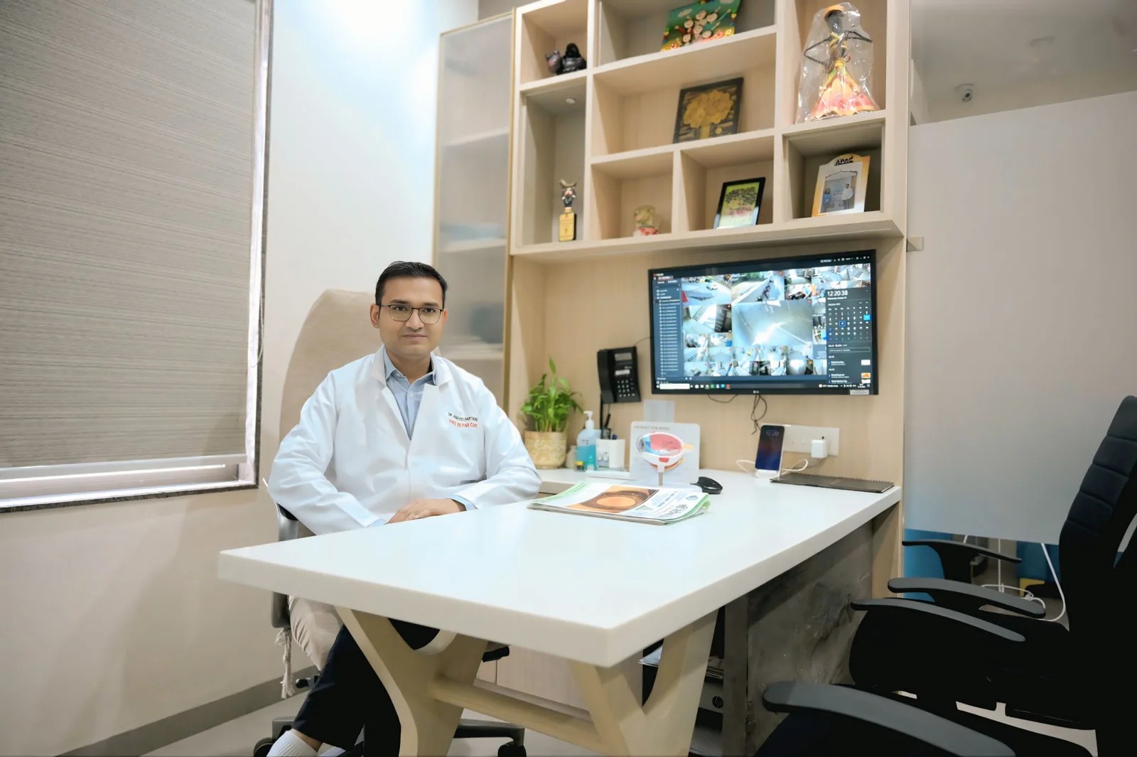
The clinic can confirm current details
The supplied clinic image is a reference, not a live price feed. Charges, test names, staff and availability can change. Call reception before travelling and request the current amount for the specific test your doctor recommended.
Shree Retina Care · TV Tower Road, Above Haldiram, Opposite Rajiv Bhawan, Shankar Nagar, Raipur, Chhattisgarh 492007.
Eye-test questions
No. The doctor decides whether OCT can help answer a question raised by the symptoms or examination. It is not a routine requirement for every visit.
Confirm the current price with reception. The supplied rate-list photo may be outdated and should not be treated as a live quote.
They use ultrasound in different ways. A-scan measures distances within the eye; B-scan forms an image of internal structures. The clinician selects a test for a specific purpose.
Yes. Clinic-supplied information lists OCT, A-scan, B-scan, FFA, pachymetry and fundus photography, among other entries. Confirm current availability and exact test names before visiting.
A test is one part of an assessment. A qualified eye-care professional interprets results with the clinical history and examination.
The materials reviewed for this page list investigations but do not confirm device manufacturers, models or technical specifications. This page does not invent those details.
Ask the clinic whether a consultation or referral is needed. A clinician can advise which investigation, if any, is useful for your concern.
This page is educational and not a substitute for professional medical advice. Test availability and charges should be verified directly with Shree Retina Care.