Keratoconus Care in Raipur: Testing and Options
Cornea health · Raipur

Keratoconus care in Raipur: get the cornea assessed

Keratoconus changes the shape and strength of the cornea, the clear front surface of the eye. It can make vision increasingly irregular, but progression and symptoms vary from person to person. A specialist assessment uses clinical examination and corneal measurements to understand what is happening and whether monitoring or treatment is appropriate.

Shree Retina Care publishes cornea-care and cornea-specialist information. The public site does not confirm a dedicated keratoconus service or a specific cross-linking schedule. Call the clinic to verify the doctor, tests and current treatment availability before travelling.

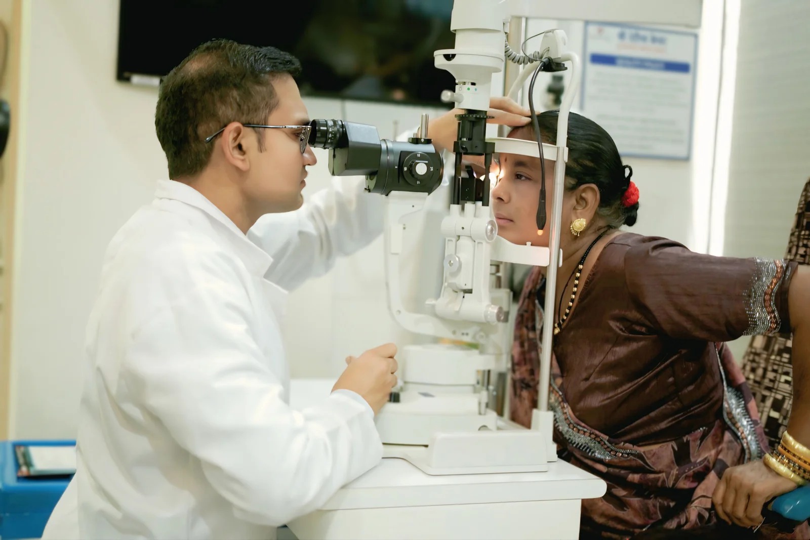
Patient receiving an eye-care consultation
Understanding the condition

What keratoconus can do to vision

The cornea normally has a smooth, regular curve. In keratoconus, the cornea becomes thinner and bulges outward in an irregular shape. That uneven surface can scatter light and create blurry, distorted or ghosted vision. A person may notice changing glasses prescriptions, increasing astigmatism, glare or difficulty seeing clearly even with an updated spectacle prescription. Symptoms can differ between the two eyes.

Keratoconus is not diagnosed from one symptom. Blurring can also come from refractive error, cataract, retinal disease, dry eye or another corneal problem. An eye examination and corneal topography or tomography can map the surface and help measure its shape. Pachymetry may be used to assess corneal thickness. A clinician compares findings with symptoms and, when appropriate, earlier scans to judge whether the condition is stable or progressing.

Family history can be relevant. Eye rubbing is associated with corneal damage and may worsen risk, so patients should avoid rubbing and ask about ways to manage itch or allergy. Do not assume rubbing alone caused the condition. A doctor can advise whether allergies, dry eye or other surface issues need treatment. Children and young adults with changing vision may need timely assessment because the cornea can change over time.

People sometimes confuse keratoconus with a cataract or assume glasses will always be enough. Glasses may help some people, especially early in the condition, but irregular corneal shape can make standard correction less effective. Other options may be considered according to vision, corneal measurements, progression and daily needs. The aim and limits of each option should be explained before a decision.

Recognize and assess

Symptoms, tests and the importance of comparison

Possible visual changes

Blur, ghost images, changing cylinder power, glare, light sensitivity and frequent prescription changes can occur. These signs are not unique to keratoconus. Sudden pain, redness or a rapid drop in vision can signal a different urgent problem and should not be attributed to keratoconus without review.

Corneal topography or tomography

Imaging maps the front surface and, depending on the device, may assess additional corneal features. It helps the doctor see irregular steepening and compare both eyes. Contact lenses can affect measurements, so the clinic may ask you to pause wearing them before a scan; follow the specific instructions supplied by the care team.

Thickness and eye examination

Pachymetry measures corneal thickness. Slit-lamp examination checks the cornea and other visible structures. The doctor also reviews vision, refraction and the health of the rest of the eye. The required tests depend on the case, so a page cannot prescribe a complete test list for every patient.

Why old scans matter

A single scan is a snapshot. When progression is being assessed, the specialist may compare reliable measurements from different dates. Differences can reflect actual change, measurement variation, contact lens effects or other factors. The clinician interprets the complete pattern rather than relying on a single number.

Do not rub your eyes. If itching or allergy makes rubbing hard to avoid, ask a doctor about safe treatment. Avoid self-prescribing steroid drops or using someone else’s contact lenses.
Management options

Treatment is selected for the person, not only the scan

The choice depends on whether the cornea is changing, how well the patient sees, the shape and thickness of the cornea, the condition of the eye surface and the patient’s age and goals. Some people need observation and updated glasses; others may need specialty contact lenses or a treatment intended to slow progression. Advanced scarring or thinning can prompt discussion of additional procedures. A doctor should explain why an option fits the findings and what it can realistically achieve.

Glasses and contact lenses

In earlier disease, spectacles may provide useful vision. As irregular astigmatism increases, rigid gas-permeable, hybrid or scleral lenses may be discussed. The appropriate lens needs fitting, instruction and follow-up; an online purchase is not a substitute. Comfort, care, hygiene and the eye’s surface must be monitored.

Corneal cross-linking

Cross-linking uses riboflavin and ultraviolet A light in a procedure intended to strengthen corneal collagen bonds and reduce the chance of further progression in selected patients. It is not designed to reshape the cornea back to normal or guarantee that glasses are no longer needed. The clinician evaluates candidacy and discusses protocol-specific risks.

Corneal rings or other options

In selected cases, an ophthalmologist may discuss intracorneal ring segments or other approaches to improve the corneal shape. Suitability depends on anatomy and history. These options have distinct benefits, risks and limitations and are not appropriate for every cornea.

Corneal transplantation

Transplant may be considered in selected advanced cases, such as when significant scarring or severe irregularity limits other options. It is not the first or inevitable treatment for everyone with keratoconus. The surgeon will explain graft type, expected recovery and long-term follow-up.

Equipment used in an eye examinationEye-care discussion with a patient and family
Questions before treatment

Understand the goal and the trade-offs

Ask the specialist to describe the purpose of the proposed care. Cross-linking is generally considered to reduce the risk of further progression; it does not promise improved unaided vision. Contact lenses can improve clarity but need fitting and ongoing care. A transplant replaces diseased tissue in selected cases but involves a longer follow-up pathway. The right choice balances disease activity, vision, risks and the patient’s preferences.

Before a procedure, clarify whether the aim is stabilization, visual improvement, surface protection or something else. Ask how the team will measure change, what result would count as success, what additional treatment may be needed and what happens if the cornea continues to change. Request the exact name of the procedure and the follow-up schedule in writing.

If cross-linking is discussed, ask which protocol is proposed and whether the cornea meets the safety criteria for that protocol. Ask about pain, bandage contact lens use, infection precautions, time away from work or school, and when the next scan is planned. Do not assume all “C3R,” “CXL” or “cross-linking” labels describe identical methods. The treating ophthalmologist should explain the method being offered.

Discuss contact lens options as well as procedures. A patient may require a lens after cross-linking or another intervention. Ask who will fit it, how frequently it should be reviewed and what to do if it becomes uncomfortable. Pain, redness or a sudden change in vision while wearing a lens should be assessed rather than managed by continuing to wear it.

For a child or teenager, ask how often vision and corneal shape need monitoring and how the plan fits school, sports and daily activities. Parents should discourage eye rubbing without blaming the child and seek help for persistent itching. Keeping a record of prescriptions and scans can make future comparisons more useful.


Living with keratoconus

Follow-up, daily habits and warning signs

Regular follow-up can help the doctor identify change and adjust treatment. The schedule depends on age, prior progression, scan findings and treatment. Attend even when vision seems stable, especially if your ophthalmologist has asked for serial corneal imaging. Bring contact lenses and a list of when they are worn if the clinician needs to assess fit or measurement effects.

Avoid eye rubbing and seek advice for allergy symptoms, dry eye or irritation. Use only prescribed eye drops. Keep contact lenses and cases clean according to professional instructions, do not sleep in lenses unless specifically instructed, and do not share lenses. Ask how often the lenses and case should be replaced. These measures support surface health but do not cure keratoconus.

Seek urgent care if vision suddenly worsens, the eye becomes painful or red, light sensitivity is severe, or a contact-lens wearer develops new pain or discharge. A sudden change is not automatically caused by keratoconus; an examination can identify the reason. Do not wait for a planned corneal scan if severe symptoms appear.

Vision needs can change as the cornea changes. Ask whether your glasses prescription, lens fit or work setup should be reviewed. If night driving, computer use or reading becomes difficult, explain the specific task. A clinician may discuss practical adjustments while continuing to monitor the cornea.

Keratoconus can affect confidence and daily routines, particularly when prescriptions change often. A clear written plan helps: the diagnosis, current scan findings, next review date, what symptoms should prompt a call and the clinic contact. If recommendations are unclear, ask for them to be restated in plain language before leaving.

Visit planning

Finding cornea care in Raipur

Shree Retina Care’s public website includes cornea-care and cornea-specialist pages and lists a Raipur clinic in Shankar Nagar. It does not publicly confirm a dedicated keratoconus clinic or the current availability of each treatment. Call before travelling to ask whether corneal topography or tomography is available, which doctor assesses keratoconus, whether you need to stop wearing contact lenses before testing and how to book follow-up imaging.

The public location is TV Tower Road, above Haldiram, opposite Rajiv Bhawan, Shankar Nagar, Raipur, Chhattisgarh 492007. Public appointment contact: +91 78980 83460; OPD contact: +91 93017 70138. Verify current numbers and doctor schedules directly. Bring old scans, glasses prescriptions, contact lens details, a medicine list and any family history you know.

Useful questions include: Is progression documented? What result supports the diagnosis? What is the aim of each proposed treatment? What are the risks in this cornea? How often should scans be repeated? What is the plan if vision changes between visits? Clear answers help patients compare options without assuming a particular intervention is required.

This page is general education, not a diagnosis or a recommendation for a procedure. Corneal imaging should be interpreted by a qualified eye-care professional in the context of a complete examination.

Patient questions

Keratoconus FAQs

Can keratoconus be cured with glasses?

Glasses can improve vision for some people, especially earlier in the condition, but they do not change the underlying corneal shape or stop progression. Other options may be discussed depending on the cornea and visual needs.

Does every person with keratoconus need C3R?

No. Cross-linking is considered for selected patients when the specialist believes the cornea is progressing and meets the relevant safety criteria. Monitoring, optical correction or another approach may be appropriate in other situations.

Does cross-linking restore normal vision?

Its main purpose is generally to reduce the chance of further progression. It may not reverse existing bulging or eliminate glasses and contact lenses. Ask the doctor to state the expected goal for your eye.

Is keratoconus the same in both eyes?

Not necessarily. The two eyes can differ in shape, severity and symptoms. Each eye should be assessed and monitored according to its own findings.

Should I stop wearing contact lenses before a corneal scan?

Contact lens wear can affect measurements. The clinic may ask you to pause lens use for a period based on the lens type. Call the testing centre for exact instructions; do not guess the time yourself.

Can eye rubbing make keratoconus worse?

Eye rubbing is associated with corneal damage and should be avoided. If allergy or irritation makes rubbing difficult to stop, ask a clinician about safe ways to manage the symptoms.

When is a cornea transplant considered?

Transplant may be discussed in selected advanced cases with significant scarring or when other options do not meet the patient’s needs. Many people never require a transplant. The decision depends on examination and corneal imaging.

Does Shree Retina Care offer keratoconus treatment?

The clinic publicly lists cornea-care information, but its public pages do not confirm a dedicated keratoconus service or specific cross-linking availability. Call to verify the doctor, tests and treatment pathway before making plans.

Further reading and review note

Sources: American Academy of Ophthalmology — Corneal Cross-Linking; AAO — Corneal Ectasia Preferred Practice Pattern; Shree Retina Care — Cornea specialist information. Confirm the exact clinic service and have an ophthalmologist review before publication. Draft prepared 23 October 2026.

Contact the clinic